Recently, the team of Zhao Xiaohang, a researcher at the Cancer Hospital of the Chinese Academy of Medical Sciences and Peking Union Medical College and at the State Key Laboratory of Molecular Oncology, together with the team of Sun Wei from the Institute of Basic Medical Sciences of the Chinese Academy of Medical Sciences, published their study titled "Noninvasive urinary protein signatures associated with colorectal cancer diagnosis and metastasis" in the internationally renowned journal Nature Communications (IF=14.919). The study conducted a discovery-verification-validation investigation using tissue samples from 993 colorectal cancer patients with a clear risk of metastasis and 657 urine samples, comprehensively identifying and validating more accurate noninvasive biomarkers in urine.
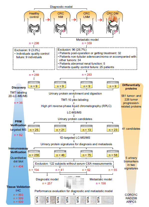
Research BackgroundColorectal cancer (CRC) is the third most common malignant tumor worldwide and the second leading cause of cancer death. It is worth noting that the clinical stage at CRC diagnosis — defined mainly by regional lymph node metastasis (LNM) and distant metastasis (DM) — is the prognostic factor most directly associated with survival and recurrence in CRC patients. In clinical practice, imaging and serum carcinoembryonic antigen (CEA) testing play a central role in monitoring CRC recurrence and metastasis. However, these mainstream imaging modalities have a number of drawbacks, including high cost, poor detection of small lymph nodes or lesions (<1 cm), and unsuitability for patients with implants or impaired renal function. Current single-modality monitoring strategies are therefore insufficient for assessing metastasis and recurrence, and more accurate and noninvasive biomarkers remain an urgent clinical need. Urine can rapidly reflect changes in the body and is a source for discovering early and sensitive biomarkers; in addition, its protein composition is markedly less complex than that of serum or plasma. Urine is therefore an excellent sample for biomarker analysis. Previous studies have identified more than 8,000 proteins in human urine; roughly 40% of urinary proteins originate from plasma proteins, and more than 1,800 proteins highly expressed in the colon can be detected in the urine of healthy individuals. It is therefore feasible to identify noninvasive biomarkers of colorectal cancer in urine. Study DesignCohort selection: CRC NM: CRC patients without any metastasis; CRC LNM: CRC patients with lymph node metastasis; CRC DM: CRC patients with distant metastasis. Healthy control: healthy individuals. Workflow: a series of MS-based and immunoassay-based methods, including a TMT-labeled quantitative proteomics strategy, a PRM-based targeted proteomics approach, quantitative dot blot analysis and tissue immunohistochemistry (Figure 1).
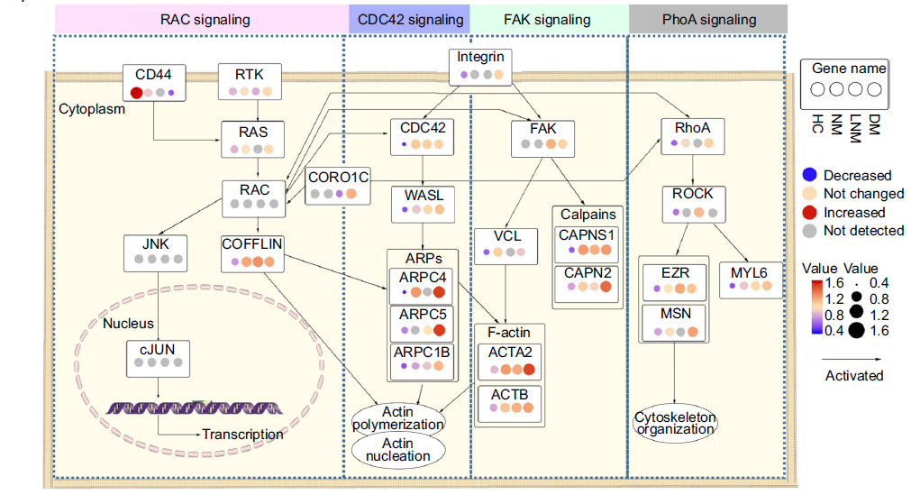
Figure 1. Overall workflow of the inclusion and exclusion criteria for study samples, and the discovery, PRM validation, immunoassay validation and tissue validation of CRC urinary biomarkers ResultsDifferential proteomic analysis of the discovery cohort Using the TMT approach, 1,976, 2,151, 2,634 and 2,771 proteins were quantified in the four groups respectively. PCA was performed on 995 proteins common to the four groups, and the results showed a clear separation between the HC and CRC groups. Pairwise differential urinary protein analyses between NM, LNM or DM and HC identified 273, 337 and 355 proteins respectively. These differential proteins were enriched in tumor-related pathways, including tumor growth, tumor invasion, immune response, metabolism and signaling pathways (RAC, FAK, CDC42 and RhoA) (Figure 2b).
|
PRM-based targeted validation
The study used PRM, a widely adopted targeted high-throughput proteomics technology, to quantitatively validate the differential urinary proteins. Among the differential proteins associated with CRC and CRC metastasis that were identified in the discovery phase, 112 and 54 proteins showing a gradient increase or decrease with CRC progression were selected for PRM validation. Of these, 77 proteins were detected in the PRM assay design and subsequently analyzed in 82 independent samples (Figure 1). Ultimately, 66 peptides from 41 differential proteins were successfully validated, with trends consistent with those obtained by the TMT approach across the four groups. Based on 23 of the upregulated proteins, a multi-level analysis was performed to define the urinary protein signature of colorectal cancer; Spearman correlation was used to evaluate them, and nine proteins moderately correlated with more than five other proteins (rho≥0.6) were excluded. The remaining 14 proteins with relatively low interdependence (median correlation coefficient of 0.34) were selected for subsequent analysis.
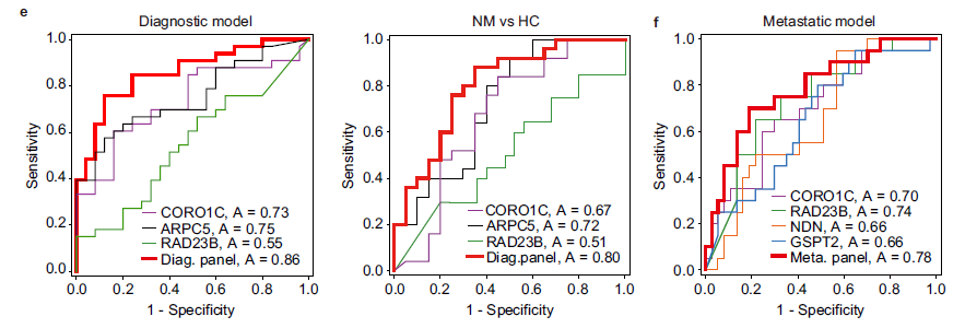
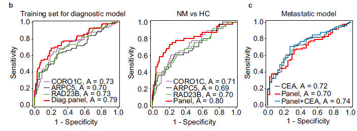
The researchers then used these 14 proteins with a random forest algorithm to identify the most important features in the diagnostic and metastasis models. ROC analysis was used at the same time to assess the classification performance of each protein in diagnosis and metastasis risk stratification. The top 10 proteins with the highest values in the two models were selected as candidate classifiers. Eight common proteins performed well in CRC diagnosis or metastasis risk stratification. In addition, the complementary performance of any two proteins was evaluated by comparing the combined AUC values for diagnosis and metastasis risk stratification. Finally, the urinary protein signature for CRC diagnosis comprised CORO1C, ARPC5 and RAD23B, while the classifier for CRC metastasis comprised CORO1C, RAD23B, GSPT2 and NDN.

Immunoassay validation of the urinary protein markers
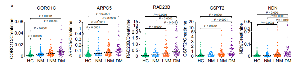
To validate the urinary signature on a large scale for distinguishing HC from CRC patients, the researchers developed a quantitative dot blot assay system using urine, based on the serum detection methods used previously. Urinary protein amounts were quantified using a standard curve for each protein and then calibrated against the corresponding urinary creatinine measurement. In the immunoassay validation phase, a total of 434 urine samples were recruited, and the results showed that the concentrations of CORO1C, ARPC5, RAD23B, GSPT2 and NDN in the urine of CRC patients were significantly higher than in HC subjects. In addition, measurements of biomarker panel fitting for CRC screening were performed on 122 samples (training set: HC, n=51; CRC, n=33; validation set: HC, n=22; CRC, n=16), and the results showed that combining the urinary protein markers with serum CEA had better predictive power than CEA alone.
Tissue proteins reflect advanced CRC and poor prognosis
To assess whether the abnormal changes in the three diagnosis-related proteins in urine originated from colorectal cancer tissue, the researchers performed immunohistochemical staining across multiple tissue microarrays. CORO1C and ARPC5 were localized in the cytoplasm, whereas RAD23B was localized mainly in the cytoplasm and nucleus. In adjacent non-cancerous tissues, staining for CORO1C, RAD23B and ARPC5 was relatively weak. In contrast, positive immunostaining for CORO1C, RAD23B and ARPC5 was observed in colorectal cancer tumor tissues, showing significant upregulation compared with adjacent non-cancerous tissues (all P<0.0001).

Subsequent Kaplan–Meier survival analysis showed that CRC patients with high CORO1C protein levels had shorter recurrence-free survival (RFS) (P=0.0075), while overall survival did not differ significantly (P=0.9171). Among CRC patients, RAD23B was significantly associated with poor overall survival (P=0.0124) but not with RFS (P=0.1123). ARPC5, however, had no prognostic significance in colorectal cancer. Taken together, since these two urinary proteins are shared by both the diagnostic and the metastasis signatures, high tissue expression of CORO1C and RAD23B does appear to promote the metastatic potential of malignant cells in colorectal cancer.

In this study, urinary proteins for CRC diagnosis and prognostic prediction were systematically analyzed by combining quantitative proteomics, targeted proteomics and immunoassays. To date, this is the largest and most comprehensive study to identify noninvasive biomarkers for colorectal cancer. The results also show that the urinary proteome can comprehensively reflect the pathophysiological changes of colorectal cancer at different stages, even at an early stage.
The findings of this study provide promising urinary protein biomarkers for the reliable diagnosis and detection of colorectal cancer, whether or not combined with the FIT test, and also point to potential intervention targets for metastatic colorectal cancer. Because only a small number of early-stage CRC patients were enrolled, the accuracy of the diagnostic model for detecting stage I CRC needs further validation. In addition, external validation in larger cohorts — especially multicenter cohorts — is required before the conclusions can be generalized. Moreover, the underlying mechanisms of these biomarkers during colorectal cancer progression remain unclear and will be a direction for future research.