
Metastasis is usually realised when circulating tumour cells (CTCs) shed from the tumour and enter the bloodstream. CTCs are tumour cells that arise from tumour tissue spontaneously or under external disturbance and enter the peripheral blood circulation or body fluids. Most CTCs undergo apoptosis or are engulfed by the body's immune cells after entering the circulation, while the few that escape may develop into metastatic foci, raising the risk of death in patients with malignant tumours.
At present, researchers generally assume that CTCs are shed during the continuous growth of tumour tissue or as a result of some form of mechanical damage, but the dynamic process that determines the generation of CTCs with metastatic capacity remains unclear.
Nicola Aceto's team at ETH Zurich carried out an in-depth study of the generation and release of CTCs, and observed an unexpected phenomenon in both breast cancer patients and mouse models: the majority of spontaneous CTC intravasation events occur during sleep. The researchers confirmed that CTCs in the resting state metastasise more readily, whereas CTCs in the active state lack metastatic capacity. This work was published online in Nature on 22 June 2022, under the title "The metastatic spread of breast cancer accelerates during sleep".
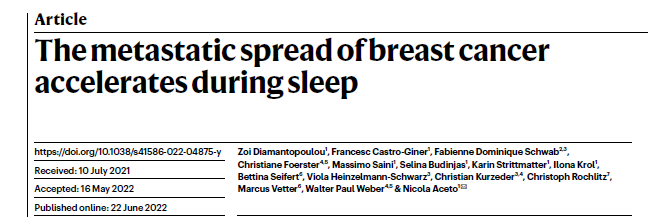
Research approach
Through single-cell RNA sequencing analysis, the researchers observed significant upregulation of mitotic genes during the rest phase in both breast cancer patients and mouse models, corroborating their metastatic capacity. The team further and systematically studied key circadian hormones such as melatonin, androgens and glucocorticoids, all of which determine the dynamics of CTC generation; insulin levels also directly promote the proliferation of tumour cells in vivo, and this process shows marked time dependence. It can therefore be concluded that CTCs with high metastatic propensity are not produced continuously, but are concentrated during the patient's sleep. This result provides a new theoretical basis for the timing control and treatment of cancers with metastatic propensity.
Circadian rhythm and the CTC release process
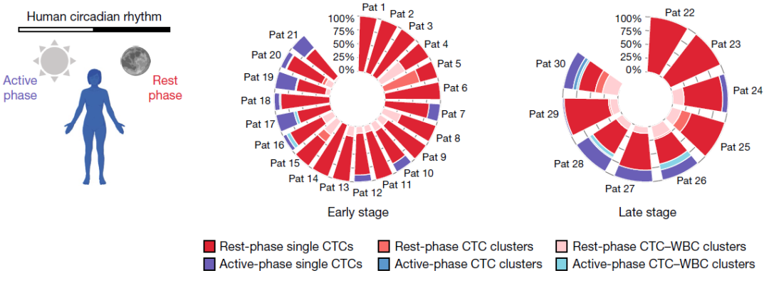
The researchers recruited 30 female patients with advanced breast cancer who were untreated or had temporarily suspended treatment — 21 patients with early breast cancer without metastatic lesions and 9 with stage IV breast cancer — and collected blood samples at an active time (10:00) and a rest time (04:00) on the same day. After capturing CTCs by microfluidic technology and performing immunofluorescence staining, the authors found that 78.3% of CTCs were collected during the rest phase, including single CTCs, CTC clusters and CTC–white blood cell clusters.
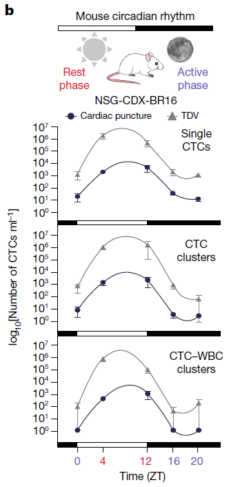
To further verify the generalisability of these findings and to describe the time dependence of their occurrence precisely, the authors used four different breast cancer mouse models: human breast CTC xenograft (NSG-CDX-BR16), human breast cancer cell xenograft (NSG-LM2), mouse breast cancer cell xenograft (NSG-4T1) and an immunocompetent syngeneic breast cancer model (BALB/c-4T1). The same phenomenon seen in the breast cancer patient data was observed in mouse blood samples, namely that most CTCs came from samples collected during the rest period. To study CTC intravasation more precisely, the researchers collected blood at 4-hour intervals over a 24-hour cycle and found a clear oscillatory pattern in CTC release, peaking between 4 and 12 hours after lights-on, that is, during the mouse rest period. Comparing CTC release at rest time (ZT4) and active time (ZT16), they found that multiple forms of CTCs were significantly increased during the rest period.
The researchers disrupted the circadian rhythm of the mice by controlling light–dark changes or by administering melatonin, a key sleep-regulating hormone. The results showed that, compared with mice on a normal circadian rhythm, CTC numbers fell significantly under a reversed day–night pattern, while melatonin induced a significant increase in CTCs. The researchers further built a single-gene knockout mouse model with rhythm disturbance; compared with wild-type mice, the circadian-rhythm-disrupted mice were unable to generate CTCs, indicating that disruption of circadian clock regulatory factors can block CTC intravasation.

Metastatic capacity of CTCs during sleep
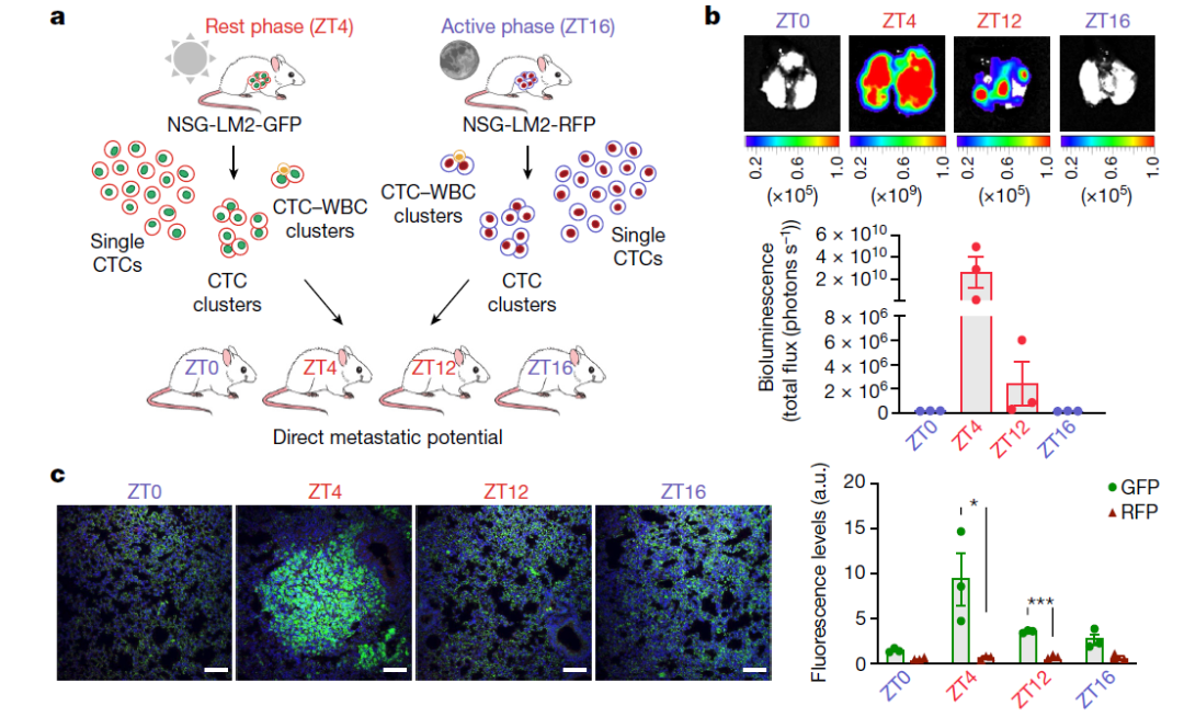
The researchers further analysed CTCs released at different circadian phases and found them to have very high metastatic capacity. They labelled CTCs collected at ZT4 in NSG-LM2 model mice with GFP and those collected at ZT16 with RFP, then mixed and injected them into the tail vein of tumour-free mice to measure metastatic capacity. In vivo bioluminescence imaging showed that the incidence of metastasis was highest during the rest period, and immunohistochemical staining confirmed that metastatic lesions mostly originated from GFP-labelled CTCs (that is, CTCs collected at ZT4). These results indicate that CTCs generated during the rest phase have higher metastatic activity and form a higher metastatic propensity during the rest phase. The researchers further injected different forms of CTCs (single CTCs, CTC clusters and CTC-WBC clusters) separately into tumour-free mice and observed their metastatic capacity, reaching the same conclusion as with mixed injection: metastatic capacity is stronger in the rest phase, and CTC clusters and CTC-WBC clusters metastasise more strongly than single CTCs. The above results show that, alongside the elevated CTC intravasation rate during the rest period, their metastatic capacity is also stronger.
Time-dependent gene expression of CTCs
The researchers went on to explore the molecular features that determine the metastatic capacity of CTCs in the rest or active phase, collecting CTCs from BR16 and LM2 mice at ZT4 and ZT16 for single-cell RNA sequencing. Principal component analysis showed that sampling time point was directly correlated with CTC gene expression levels. Comparing gene expression between samples collected at ZT4 and ZT16, a total of 121 genes were upregulated at ZT4 and 156 at ZT16. The trends of characteristic gene changes were consistent across different CTC forms, with the greatest statistical differences in CTC clusters and CTC-WBC clusters. Gene set enrichment analysis (GSEA) showed that the upregulated genes in CTCs isolated at ZT4 were mainly related to mitosis and cell division pathways, while those upregulated in CTCs isolated at ZT16 were mainly related to ribosome biogenesis and gene translation pathways — findings consistent with the typical temporal patterns of gene expression in eukaryotic cells. Staining for the tumour proliferation marker Ki67 also showed significant upregulation during the rest phase, consistent with the changes in CTC intravasation and mitotic gene expression. This periodic fluctuation appeared not only in CTCs but also in the primary tumour, indicating that it is a general phenomenon in breast cancer cells during disease progression.
Regulators of the CTC intravasation process
Finally, the researchers investigated the main regulators by which the circadian rhythm drives CTC generation and proliferation. First, by analysing the differential expression of canonical clock genes in cancer cells between the rest and active phases, no significant difference was detected in either CTCs or primary tumour cells, although rhythmic changes could be observed in non-tumour tissue. Further analysis of the relationship between oscillatory changes in CTC counts and factors such as interstitial fluid pressure, immune cell interaction or haemolysis rate likewise revealed no significant rhythmic difference. The researchers then analysed the receptor expression levels of 63 known circadian-regulated hormones and growth factors in RNA sequencing data from patients and mouse models. The expression of glucocorticoid receptor, androgen receptor and insulin receptor was high across all forms of CTCs, indicating that these ligands participate in the time-rhythmic generation and proliferation of CTCs.
Summary
Previous studies have demonstrated the role of the circadian rhythm in tumorigenesis, but the dynamics that prospectively regulate the metastatic progression process remain unclear. This study addresses the generation of CTCs with metastatic capacity, showing in both breast cancer patients and mouse models that CTC generation is concentrated in the resting state and carries a higher metastatic propensity. This enhanced metastatic capacity derives from time-dependent changes in proliferation rate and is influenced by circadian-regulating hormones. The results indicate that control of circadian timing is critical to the study and treatment of breast cancer.
