Recently, a team from the Second Xiangya Hospital of Central South University and its research institute published an article in the Chinese Journal of Laboratory Medicine titled "Investigating the cause of the postprandial decline in low-density lipoprotein cholesterol measured by the direct method." Based on NMR (nuclear magnetic resonance) spectroscopy, the article examines why postprandial LDL-C measured by the direct method falls below fasting levels, and concludes that postprandial LDL-C obtained by the direct method is not suitable for assessing the risk of cardiovascular events. The authors give special thanks to ProteinT for its support in NMR testing. Relying on a powerful NMR platform, ProteinT continues to explore early warning, medication guidance and long-term monitoring for cardiovascular and cerebrovascular diseases, advancing more precise screening and medication guidance for conditions including cardiovascular and cerebrovascular diseases and common cancer types.
Background
Disordered postprandial lipid metabolism is a risk factor for coronary heart disease, and LDL-C falls markedly after eating. Some guidelines therefore use non-fasting lipid levels to predict the risk of coronary heart disease. Whether this decline reflects an absolute reduction in cholesterol or a redistribution of cholesterol, however, remains unclear.
In clinical practice, the direct method is more commonly used to measure LDL-C, but it cannot precisely detect lipoprotein subfractions and its results are influenced by changes in lipoprotein subfraction particle size. Recent studies have found that nuclear magnetic resonance spectroscopy (NMRS) can measure the number, density, size and composition of lipoproteins, and that its results are less readily affected by changes in lipoprotein composition.
This study set out to compare postprandial lipid measurements obtained by the direct method and by NMRS and to investigate changes in postprandial lipid subfractions, helping to identify the true cause of the postprandial decline in LDL-C. It also measured postprandial PCSK9 levels to explore their relationship with lipid changes.
Methods
1. Study population
The study enrolled 85 subjects at the Second Xiangya Hospital of Central South University between November 2017 and May 2019.
2. Grouping
Subjects were divided into a coronary heart disease (CHD) group (n=33) and a non-CHD group (n=52). The non-CHD group was further divided into a high-TG group (n=13) and a normal group (n=39). The CHD group comprised patients with ischemic electrocardiographic abnormalities whose angiography showed stenosis of at least 50% in one or more coronary arteries. The high-TG group comprised patients with fasting TG>2.26 mmol/L. The normal group comprised subjects with neither high TG nor CHD, excluding patients with renal failure, malignant tumors and other conditions.
3. Laboratory tests and NMR spectroscopic lipid measurement
Clinical information was recorded for all subjects. Blood samples were taken in the fasting state and at 2 h and 4 h after a meal; serum was separated and stored at −80 °C. Biochemical and lipid indices were measured by the direct method; lipoprotein particles and their subfractions were measured by NMR spectroscopy; and serum PCSK9 levels were determined by enzyme-linked immunosorbent assay.
4. Statistical analysis
Normally distributed data were analyzed by one-way analysis of variance, with the Bonferroni method used for pairwise comparisons; non-normally distributed data were analyzed by Friedman's two-way analysis of variance by ranks, with the Wilcoxon signed-rank test used for pairwise comparisons. Categorical variables were analyzed using the χ² test. Correlations between PCSK9 levels and lipoprotein particle numbers were assessed by Spearman correlation analysis. P< 0.05 was considered statistically significant.
Study design
In all patients, as well as in patients with coronary heart disease, diabetes mellitus (DM) and metabolic syndrome (MS), multivariate logistic regression and Spearman correlation analysis were used to explore the risk factors for coronary heart disease and the relationships between the Gensini score, PCSK9 and NMR results.
Results
1. Baseline characteristics
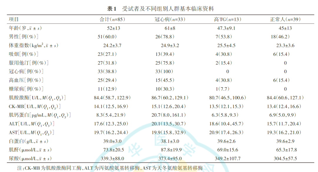
A total of 85 subjects were enrolled; coronary heart disease, hypertension and diabetes accounted for 38.8%, 29.4% and 12.9% respectively, and 31.8% of patients were taking statins. Details are shown in Table 1.
2. Changes in cholesterol within postprandial atherogenic lipoprotein particles
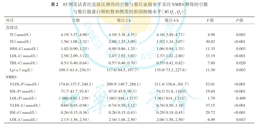
Postprandial lipid measurements obtained by the direct method and by NMRS in the 85 subjects are shown in Table 2. Compared with the fasting state, LDL-C concentrations measured by the direct method decreased at 2 h and 4 h after the meal (P<0.001), while TRL-C increased (P=0.020). Differences in LDL-C concentration measured by NMRS were statistically significant (P=0.047); postprandial TRL-C rose (P=0.015); and both VLDL-C and IDL-C increased at 4 h postprandially (P<0.001).
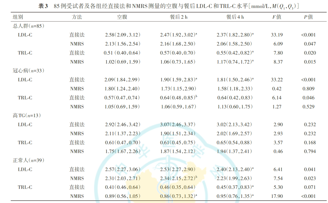
Fasting and postprandial LDL-C and TRL-C levels measured by the direct method and by NMRS in all 85 subjects and in each group are shown in Table 3. Among the 13 subjects with high TG, neither the direct method nor NMRS showed a statistically significant difference in postprandial LDL-C (P=0.232); among the 33 subjects with CHD, the direct method showed a postprandial decrease in LDL-C (P<0.001), while the NMRS result was not statistically significant (P=0.809); among the 39 normal subjects, the direct method showed a postprandial decrease in LDL-C (P=0.041) and NMRS showed a statistically significant difference in postprandial LDL-C (P=0.023).
3. NMRS measurement of changes in postprandial LDL subfractions
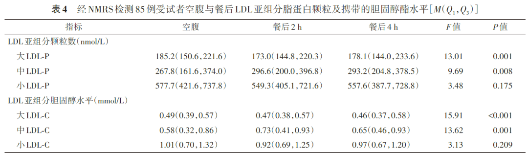
The concentrations of the different postprandial LDL subfraction particles changed: large LDL-P concentration decreased (P=0.001) and medium LDL-P concentration increased (P=0.008). Postprandial cholesterol was redistributed among the different LDL subfractions: large LDL-C concentration fell (P<0.001) and medium LDL-C concentration rose (P=0.001). Differences in small LDL-P and LDL-C concentrations were not statistically significant (P>0.05). Details are shown in Table 4.
4. Relationship between the postprandial decrease in PCSK9 and the postprandial decrease in LDL-C and increase in remnant cholesterol (RC)
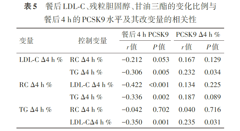
PCSK9 levels in the fasting state and at 2 h and 4 h postprandially were 299 (233, 397), 257 (208, 342) and 251 (215, 340) ng/ml respectively, a statistically significant difference (P<0.001). The change in PCSK9 at 4 h postprandially was positively correlated with the change in LDL-C measured by the direct method (r=0.172, P=0.020), but no correlation remained after adjustment for RC (r=0.167, P=0.129, Table 5).
NMRS results showed that after adjusting for LDL-C and TG respectively, the change in RC at 4 h postprandially was negatively correlated with PCSK9 levels (r=−0.422, P<0.001; r=−0.336, P=0.002, Table 5).
The magnitude of the postprandial change in RC measured by NMRS was positively correlated with the decrease in PCSK9 levels (r=0.232, P=0.035).
Discussion
Although the direct method shows a marked postprandial reduction in LDL-C in clinical practice, NMRS did not observe a reduction in LDL particles. This indicates that after a meal the cholesterol in LDL is not cleared by the liver, but is redistributed among the different LDL subfractions, mainly between large and medium LDL particles. Direct-method LDL-C may therefore reflect the cholesterol concentration in large LDL particles more than the cholesterol concentration across all LDL.
Postprandial serum PCSK9 levels fell, and this was not correlated with the decrease in LDL particle number or LDL-C. PCSK9 levels are likewise influenced by the postprandial influx of cholesterol into the liver: this study found that the proportion by which postprandial PCSK9 fell was negatively correlated with the rate at which RC increased, indicating that the postprandial decline in serum PCSK9 is caused by food intake. The study also observed that the increase in RC at 4 h postprandially was negatively correlated with PCSK9, indicating that the postprandial reduction in PCSK9 is a compensatory response of intrahepatic cholesterol homeostasis.
Summary
The transfer of cholesterol from large LDL particles into medium and small LDL particles lowers and redistributes LDL-C, making LDL more atherogenic; the rise in intrahepatic cholesterol concentration after a meal may lead to a fall in postprandial serum PCSK9 levels. Postprandial LDL-C measured by the direct method is therefore not suitable for assessing the risk of cardiovascular events.

