
Recently, the cardiology team at the Second Xiangya Hospital of Central South University published an article in the Chinese Journal of Cardiovascular Medicine (online) titled "High-sensitivity C-reactive protein may be a biomarker of high-density lipoprotein dysfunction and structural remodeling in patients with coronary atherosclerotic heart disease." Based on nuclear magnetic resonance (NMR) spectroscopy, the study explored indicators in patients with coronary heart disease that reflect the cholesterol efflux capacity (CEC) mediated by high-density lipoprotein (HDL). The authors specifically acknowledged ProteinT for its support in NMR testing and analysis. Building on the advantages of NMR spectroscopy—non-destructive testing with minimal sample volumes, fully automated high throughput, unparalleled reproducibility and a proprietary database platform—ProteinT continues to explore and investigate early warning, medication guidance and long-term monitoring in cardiovascular and cerebrovascular diseases, advancing more precise screening and treatment guidance for cardiovascular and cerebrovascular conditions as well as common cancers.
Background
The Global Burden of Disease study and the Report on Cardiovascular Health and Diseases in China show that atherosclerotic cardiovascular disease, represented by coronary atherosclerotic heart disease (CHD), remains the leading cause of death worldwide, and that CHD mortality in China is still rising year by year—making prevention and treatment urgently necessary. Although current therapies that lower low-density lipoprotein cholesterol (LDL-C) have produced significant benefits in the prevention and treatment of CHD, about 10% of patients still experience cardiovascular events even when LDL-C is reduced to as low as 0.78 mmol/L. This residual risk urges us to keep actively searching for new intervention targets. In the 1980s, a number of epidemiological studies consistently found that high-density lipoprotein cholesterol (HDL-C) levels were inversely associated with the incidence of CHD events, and HDL-C was therefore regarded as an important cardiovascular protective factor. Subsequent clinical trials of drugs that raise HDL-C and genetic studies, however, did not support this view. HDL is a highly heterogeneous group of lipoprotein particles 8–13 nm in diameter in plasma, with functions including mediating cholesterol efflux from foam cells, antioxidation and anti-inflammatory effects on endothelial cells. At present, multiple large-scale clinical trials have confirmed that HDL cholesterol efflux capacity (CEC), measured in vitro, can independently predict cardiovascular risk in healthy populations as well as the incidence of adverse events and mortality in patients with CHD, giving it important clinical value. However, measuring CEC requires in vitro cell models and isotopes, which limits its use in routine clinical practice. In this study, we therefore sought to identify indicators that can reflect CEC in patients with CHD.
Methods
We consecutively enrolled 36 patients with coronary heart disease (CHD group) who were hospitalized in the Department of Cardiology at the Second Xiangya Hospital of Central South University between June and July 2018 and confirmed by coronary angiography, together with 61 patients without coronary heart disease (non-CHD group) over the same period. Fasting blood was collected the morning after admission, and plasma samples were obtained after centrifugation of whole blood. Plasma samples were aliquoted and stored frozen at -80 °C for CEC and HDL structure testing. CEC measurement involved five main steps: obtaining HDL by heparin-manganese precipitation, loading macrophages with lipid, up-regulating the expression of cholesterol efflux channel proteins, cholesterol efflux, and detecting isotope content. HDL structure was measured using nuclear magnetic resonance spectroscopy (NMRS). Baseline characteristics and routine biochemical indicators of the subjects were collected, and CEC and HDL structural parameters were measured. Pearson correlation analysis was used to identify variables associated with CEC, and multiple linear regression analysis was used to further evaluate the relationship between these variables and CEC.
Results
1. Clinical characteristics
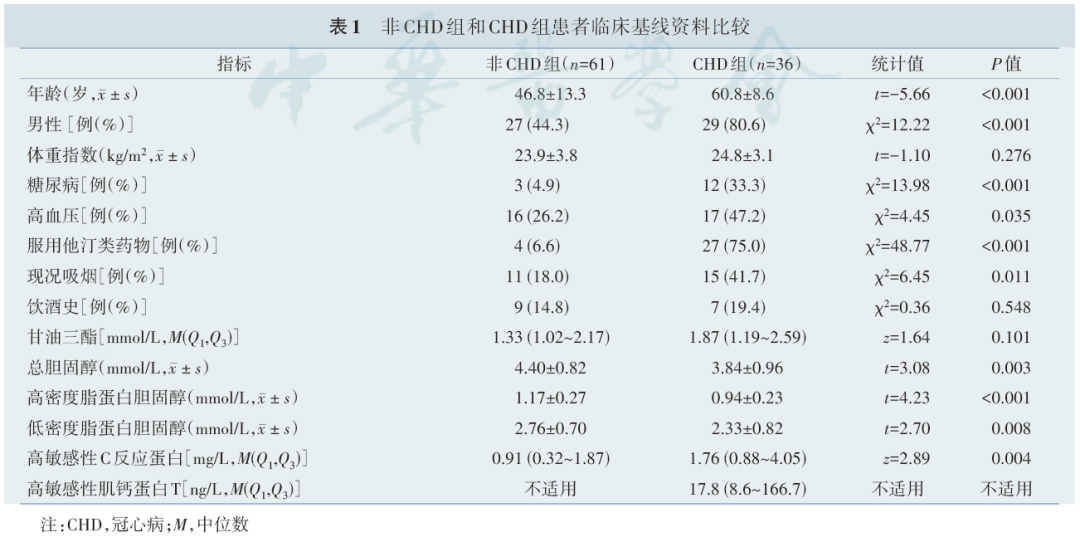
All patients in the CHD group were diagnosed with acute coronary syndrome, including 4 cases of ST-segment elevation myocardial infarction, 14 cases of non-ST-segment elevation myocardial infarction and 18 cases of unstable angina. Among them, blood samples from 29 patients (80.6%) were collected at least 7 days after onset. Compared with the non-CHD group, patients in the CHD group were older and included more men, as well as more cases of diabetes, hypertension and current smoking; plasma total cholesterol, HDL-C and LDL-C were lower, while hs-CRP was higher [1.76 (0.88–4.05) vs. 0.91 (0.32–1.87), Z=2.89, P=0.004].
2. Relationship between HDL cholesterol and cholesterol efflux capacity
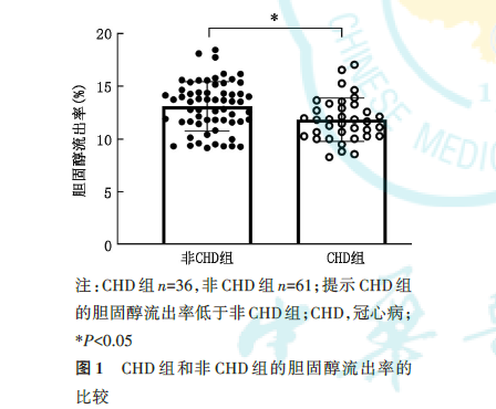
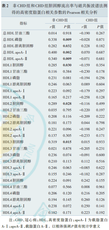
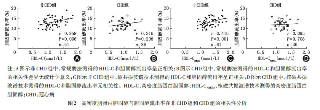
CEC in the CHD group was lower than in the non-CHD group [(11.9%±2.3%) vs. (13.0%±2.2%), t=-2.32, P=0.022, Figure 1], a statistically significant difference. In the non-CAD group, HDL-C was positively correlated with CEC (r=0.358, P=0.006, Figure 2A), whereas this correlation was not statistically significant in the CAD group (r=0.216, P=0.206, Figure 2B). NMRS confirmed this result: HDL-C NMRS was positively correlated with CEC in the non-CHD group (r=0.416, P=0.001, Figure 2C), but showed no correlation with CEC in the CAD group (r=0.065, P=0.708, Figure 2D). NMRS also measured the contents of apolipoprotein A-I (apoA-I), apoA-II, phospholipids, triglycerides and free cholesterol in total HDL and in each HDL subgroup. Univariate correlation analysis between CEC and the above indicators showed that in the non-CHD group, CEC was positively correlated with total HDL phospholipids, total HDL free cholesterol, total HDL apoA-I and total HDL apoA-II (P<0.05), whereas in the CHD group all of these correlations disappeared.
3. Factors influencing cholesterol efflux capacity in the coronary heart disease group
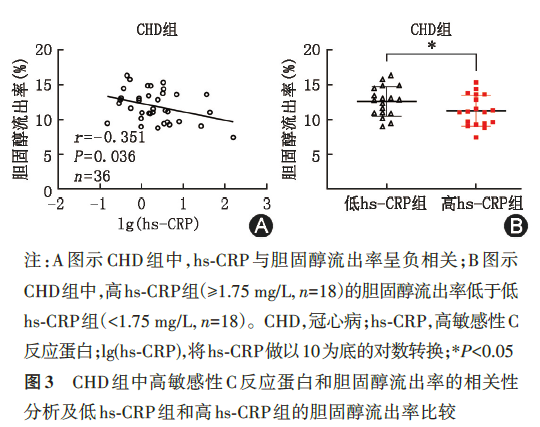
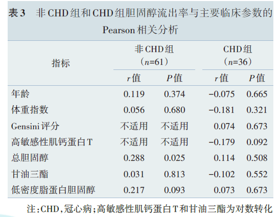
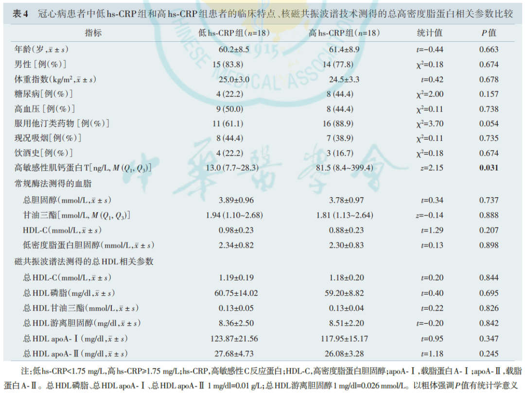
To explore the factors influencing CEC in the CHD group, we conducted correlation analysis between various clinical indicators and CEC. CEC showed no correlation with patient age, body mass index, severity of coronary lesions (Gensini score), high-sensitivity troponin T, total cholesterol, triglycerides or LDL cholesterol (Table 3). However, CEC was negatively correlated with hs-CRP (r=-0.351, P=0.036, Figure 3A). The 36 CAD patients were divided into a high hs-CRP group and a low hs-CRP group according to the median hs-CRP (1.75 mg/L). Apart from high-sensitivity troponin T, there were no significant differences between the two groups in baseline characteristics or in the lipid and apolipoprotein contents of total HDL (Table 4), but CEC was lower in the high hs-CRP group than in the low hs-CRP group [(11.3%±2.2%) vs. (12.6%±2.1%), t=-1.83, P=0.038, Figure 3B], a statistically significant difference.
4. Composition of HDL in the coronary heart disease group
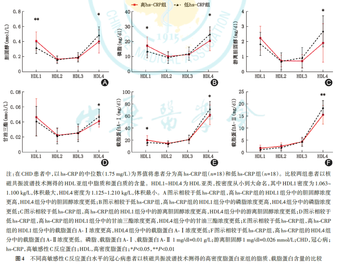
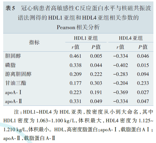
NMRS was used to examine HDL structure in CHD patients with different hs-CRP levels. The results showed that, compared with the low hs-CRP group, the high hs-CRP group had higher lipid and apolipoprotein contents in the largest HDL subgroup (HDL1) and lower lipid and apolipoprotein contents in the smallest HDL subgroup (HDL4) (Figure 4). Correlation analysis showed that hs-CRP was positively correlated with the lipid and apolipoprotein contents of HDL1 and negatively correlated with those of HDL4.
5. Multiple linear regression analysis
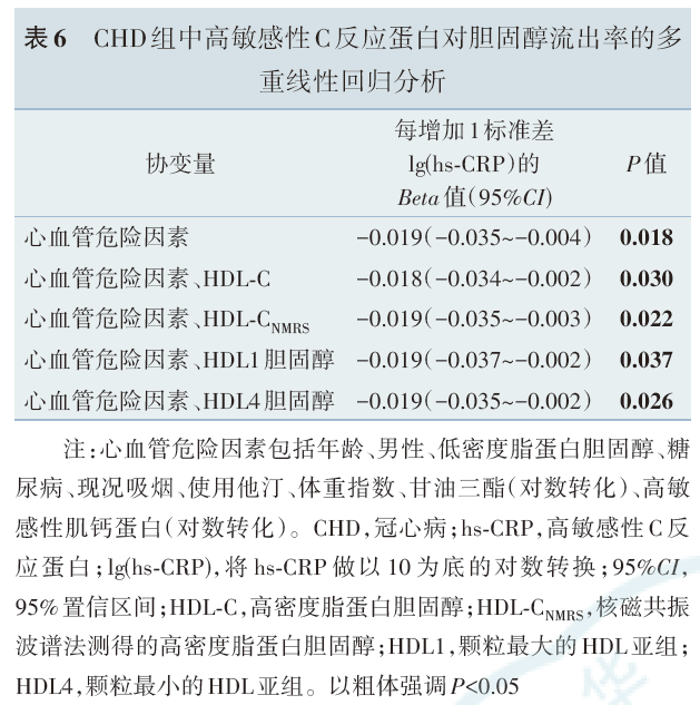
In the CHD group, after adjusting for age, male sex, LDL cholesterol, diabetes, current smoking, body mass index, triglycerides, high-sensitivity troponin and statin use, and then separately controlling for HDL-C / HDL-C NMRS / HDL1 cholesterol / HDL4 cholesterol, hs-CRP remained negatively correlated with CEC.
Discussion
For a long time, HDL-C was widely accepted as a protective factor against atherosclerotic cardiovascular disease, yet most clinical trials of drugs aimed at raising HDL-C—such as niacin and cholesteryl ester transfer protein inhibitors—ended in failure. More recently, an epidemiological study of 43,407 healthy individuals with a mean follow-up of 12.1 years found that very high HDL-C levels (≥2.32 mmol/L) were associated with an increased risk of cardiovascular death. This shows that predicting cardiovascular risk from HDL-C levels alone is unreliable. HDL exerts its anti-atherosclerotic effect mainly by mediating the reverse transport of cholesterol from beneath the vascular endothelium to the liver. CEC is an in vitro measure that can be used to evaluate the initial step of reverse cholesterol transport, namely HDL-mediated removal of cholesterol from cells. Multiple studies have established CEC as an independent protective factor against cardiovascular events. However, measuring CEC places high demands on the laboratory environment and is time- and labor-intensive. How to evaluate CEC easily in patients with coronary heart disease is therefore a question worth exploring.
In this study, we explored HDL function and structure in small samples of non-CHD and CHD patients. The findings were as follows: (1) CEC was lower in the CHD group than in the non-CHD group [(11.9%±2.3%) vs. (13.0%±2.2%)], a statistically significant difference. In the non-CHD group, CEC was positively correlated with HDL-C (enzymatic method: r=0.358, P=0.006; NMRS: r=0.416, P=0.001), whereas in the CHD group there was no correlation between CEC and HDL-C (enzymatic method: r=0.216, P=0.206; NMRS: r=0.065, P=0.708); (2) in the CHD group, CEC was negatively correlated with hs-CRP (r=-0.351, P=0.036), and this correlation persisted after adjustment for CHD risk factors and HDL-C, indicating that hs-CRP is a risk factor for reduced CEC; (3) in the CHD group, the higher the hs-CRP, the more large HDL particles and the fewer small HDL particles, indicating remodeling of HDL toward larger particles.
1. Reduced HDL-C efflux capacity in patients with coronary heart disease
High-quality previous studies have found that CEC is significantly lower in patients with coronary heart disease than in healthy individuals, and our results are consistent with these. The literature, however, remains controversial regarding the relationship between HDL-C and CEC in CHD patients. The correlation coefficient between HDL-C and CEC was 0.51 (P<0.0001) in the study by Khera et al., but -0.09 in the study by Zhang et al. Our results agree with those of Zhang et al., possibly because both our study and that of Zhang et al. enrolled patients with acute coronary syndrome, whereas Khera et al. enrolled patients with stable coronary heart disease together with some healthy individuals. It follows that disease state influences CEC. Besides coronary heart disease, impaired CEC is also seen in patients with acute inflammation, end-stage renal disease, type 1/2 diabetes and autoimmune diseases.
2. In patients with coronary heart disease, cholesterol efflux capacity is negatively correlated with high-sensitivity C-reactive protein
We also observed that in patients with coronary heart disease, the decline in CEC was accompanied by a negative correlation between hs-CRP and CEC. This phenomenon closely resembles that found by Vaisar et al. in an acute inflammation model. In that study, a low dose of endotoxin was used to induce an acute inflammatory response in healthy subjects, and CEC declined. HDL proteomics at that point revealed marked increases in serum amyloid A1 (SAA1) and SAA2 within HDL, with SAA negatively correlated with CEC. Although the cause has not been fully elucidated, in vitro experiments and SAA1/2 knockout mouse models confirmed that SAA incorporated into HDL can itself cause CEC to decline. Because coronary heart disease is also an inflammatory disease, and because CRP, like SAA, is an acute-phase protein secreted by the liver during systemic inflammatory responses, we speculate that the decline in CEC and its negative correlation with hs-CRP observed in our study may result from increased SAA within HDL. On the other hand, inflammatory apoA-I modification can also impair CEC. In this study, total HDL apoA-I was correlated with CEC in healthy individuals, whereas this correlation disappeared in patients with coronary heart disease, suggesting that inflammatory apoA-I modification may also be one of the causes of the phenomenon described above. Although the mechanisms of inflammation in the development and progression of coronary heart disease have not been fully clarified, hs-CRP is already an established risk marker for CHD. Patients with stable coronary heart disease and high baseline hs-CRP (≥2 mg/L) have a significantly higher risk of major adverse cardiovascular events (MACE) than those with low baseline hs-CRP. In the CANTOS trial, patients with prior myocardial infarction and hs-CRP ≥2 mg/L were treated with canakinumab, a monoclonal antibody against interleukin-1β. Among those whose hs-CRP fell below 2 mg/L during treatment, MACE events were reduced by 25%, whereas those who did not reach the hs-CRP target derived no significant benefit. By contrast, another anti-inflammatory drug, methotrexate, conferred no benefit in patients with prior myocardial infarction, accompanied by no significant reduction in hs-CRP. Based on the above findings and those of our study, one reason—or at least one reason—why hs-CRP can serve as a risk marker for coronary heart disease is that it reflects HDL dysfunction.
3. HDL is remodeled toward larger particles in CHD patients with high hs-CRP levels
We also found that in CHD patients with high hs-CRP levels, plasma HDL was remodeled toward larger particles. This is consistent with previous results obtained by separating HDL subclasses using electrophoresis. Mature HDL is a spherical particle formed from a hydrophobic lipid core (mainly cholesteryl esters and triglycerides) surrounded by a monolayer of phospholipids, with apolipoproteins on its surface. Based on particle size, HDL can be divided into large and small particles. Epidemiological studies have found that in healthy populations, small HDL particles are negatively correlated with the incidence of CHD, whereas large particles show no clear correlation. This study did not investigate the mechanism of HDL remodeling in depth, but it is worth noting that the incorporation of SAA described above can also produce a dose-dependent increase in HDL size.
4. Limitations of this study
This study has certain limitations. First, because the samples and data came from another cross-sectional study whose original purpose was not to find biomarkers of CEC, the sample size of CHD patients was relatively small. Second, in the multiple linear regression analysis, although hs-CRP remained negatively correlated with CEC after adjustment for cardiovascular risk factors (P<0.05), if the P value were corrected using the Bonferroni test, the correlation would lose statistical significance; we can therefore only conclude that the two are "possibly correlated," and further studies are needed to reach a definitive conclusion. Third, half of the patients with coronary heart disease enrolled in this study were diagnosed with myocardial infarction. The source of inflammation in patients with myocardial infarction (mainly necrotic myocardium) differs from that in patients with stable coronary heart disease (mainly the vessel wall or adipose tissue), and the mechanisms by which they affect HDL function may differ. Large-scale clinical studies are still needed to separately investigate HDL structure and function in patients with stable coronary heart disease and in patients in the acute phase of myocardial infarction, in order to further verify these conclusions. Finally, although this study speculates that systemic inflammation plays an important role in HDL structural and functional remodeling, we did not investigate its mechanism in depth.
Conclusions
In summary, in patients with coronary heart disease, HDL-C does not reflect HDL's CEC, whereas measuring hs-CRP can. The higher the hs-CRP level, the lower the HDL CEC, accompanied by remodeling of HDL toward larger particles. hs-CRP may be a biomarker of HDL dysfunction and structural remodeling in patients with coronary heart disease, and systemic inflammation may be the common mechanism underlying these phenomena.
